Home
/ Anatomy Of Back Of Neck, Second Layer Of Back And Neck Muscles 1866 Illustration Stock Image C040 5297 Science Photo Library : The neck is the area between the skull base and the clavicles.
Anatomy Of Back Of Neck, Second Layer Of Back And Neck Muscles 1866 Illustration Stock Image C040 5297 Science Photo Library : The neck is the area between the skull base and the clavicles.
Anatomy Of Back Of Neck, Second Layer Of Back And Neck Muscles 1866 Illustration Stock Image C040 5297 Science Photo Library : The neck is the area between the skull base and the clavicles.. Learn about the various causes of back pain, including different kinds of arthritis. Anatomy of the hand overview. Most of the problems or conditions from the neck come from the bottom 5 vertebra (cervical vertebra 3 through cervical vertebra 7 or more anatomy of lumbar spine. A dynamic and interactive atlas of ent imaging. The physicians originally studying human anatomy thought the skull looked like an helmet.
Despite being a relatively small region, it contains a range of important anatomical features. Your neck is like no other part of the vertebral spinal column and enables your head and neck a wide range of motion. Want to learn more about it? This article looks at the anatomy of the back, including bones, muscles, and nerves. Join our newsletter and receive our free ebook:
The Anatomy Of The Neck And Back Of The Head Posters Prints By Unknown from previews.magnoliabox.com Learn more about head and neck anatomy, including the top part of the skeleton, muscles, and more with our digital flashcards. From the sides and the back of the neck, the splenius capitis inserts onto the head region, and the splenius. The head rests on the top part of the vertebral column, with the skull joining at c1. The splenius muscles originate at the midline and run laterally and superiorly to their insertions. Despite being a relatively small region, it contains a range of important anatomical features. The head and neck meet at the superior nuchal line. The neck is the area between the skull base and the clavicles. The neck muscles, including the sternocleidomastoid and the trapezius, are responsible for the gross motor movement in the muscular system of the head and neck.
This human anatomy lecture has been made by wendy riggs, for the college of the.
Despite being a relatively small region, it contains a range of important anatomical features. The splenius muscles originate at the midline and run laterally and superiorly to their insertions. Head and neck anatomy is important when considering pathology affecting the same area. The splenius muscles originate at the midline and run laterally and superiorly to their insertions. Clinically, surface anatomy is used to split the neck into anterior and posterior triangles which provide clues as to the location of specific structures. A coronal and axial contrast enhanced multidetector computed tomography imaging of the head and neck was performed on a healthy subject. It is made up of bones discs muscles ligaments nerves and tendons. By understanding the anatomy of the neck and how each structure works, it's easier to understand the sources of neck pain. The cervical spine supports the weight and movement of your head and pro. The arteries that ultimately supply the head and neck originate from the subclavian and common carotid arteries. Many conditions and injuries can affect the back. Most of the problems or conditions from the neck come from the bottom 5 vertebra (cervical vertebra 3 through cervical vertebra 7 or more anatomy of lumbar spine. Guide to mastering the study of anatomy.
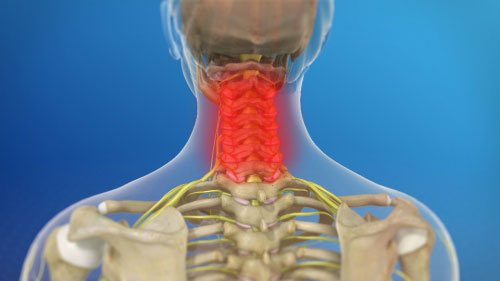
The neck muscles, including the sternocleidomastoid and the trapezius, are responsible for the gross motor movement in the muscular system of the head and neck. Learn everything about the neck anatomy with this topic page. From the sides and the back of the neck, the splenius capitis inserts onto the head region, and the splenius. The cervical spine supports the weight and movement of your head and protects the nerves exiting your brain. Most of the problems or conditions from the neck come from the bottom 5 vertebra (cervical vertebra 3 through cervical vertebra 7 or more anatomy of lumbar spine.
Neck Atlas Of Anatomy from doctorlib.info The cervical spine supports the weight and movement of your head and pro. 3d video tutorials and interactive modules on the anatomy of the back including anatomy of the musculature, vertebral column, joints and ligaments. This article looks at the anatomy of the back, including bones, muscles, and nerves. The neck, cheek (lower face), temple and the scalp are arbitrarily divided by the lower border of the mandible, the zygomatic arch and the temporal line, respectively. We've largely focused on the physical aspect of our spinal anatomy in this series. Its midpoint is marked by the external occipital protuberance. From the sides and the back of the neck, the splenius capitis inserts onto the head region, and the splenius. Additionally, the joints in the back of the cervical vertebrae (facets) are shaped to allow movement:
The cervical spine supports the weight and movement of your head and pro.
Most of the problems or conditions from the neck come from the bottom 5 vertebra (cervical vertebra 3 through cervical vertebra 7 or more anatomy of lumbar spine. The arteries that ultimately supply the head and neck originate from the subclavian and common carotid arteries. From the sides and the back of the neck, the splenius capitis inserts onto the head region, and the splenius. Our neck is where we find the seven cervical vertebrae, with c7 (the seventh cervical vertebra) meeting t1 (the first thoracic vertebra) at the base of the neck. The physicians originally studying human anatomy thought the skull looked like an helmet. Learn about the various causes of back pain, including different kinds of arthritis. Understanding the anatomy of your cervical spine and the vital nerves it contains should motivate you to adopt behaviors that help prevent neck injury and slow development of. Resists back hyperextension, c1 to sacrum resists hyperflexion of the back, helps prevent herniation, c2… Demonstrate sound knowledge of the surface/living and radiological anatomy of the head, neck and. The physicians originally studying human anatomy thought the skull looked like an apple. From the sides and the back of the neck, the splenius capitis inserts onto the head region, and the splenius cervicis extends onto the cervical region. Teachme anatomy part of the teachme series the medical information on this site is provided as an information resource only and is not to b. Guide to mastering the study of anatomy.
A collection of anatomy notes covering the key anatomy concepts that medical students need to learn. Resists back hyperextension, c1 to sacrum resists hyperflexion of the back, helps prevent herniation, c2… It is made up of bones discs muscles ligaments nerves and tendons. From the sides and the back of the neck, the splenius capitis inserts onto the head region, and the splenius cervicis extends onto the cervical region. Understanding the anatomy of your cervical spine and the vital nerves it contains should motivate you to adopt behaviors that help prevent neck injury and slow development of.
Tips For Weekend Warriors Preventing Back And Neck Injuries Baldwin Bone Joint Pc from baldwinboneandjoint.com The arteries that ultimately supply the head and neck originate from the subclavian and common carotid arteries. Most of the problems or conditions from the neck come from the bottom 5 vertebra (cervical vertebra 3 through cervical vertebra 7 or more anatomy of lumbar spine. A collection of anatomy notes covering the key anatomy concepts that medical students need to learn. Join our newsletter and receive our free ebook: Teachme anatomy part of the teachme series the medical information on this site is provided as an information resource only and is not to b. Neck, in land vertebrates, the portion of the body joining the head to the shoulders and chest. The head and neck meet at the superior nuchal line. A dynamic and interactive atlas of ent imaging.
In the neck, the platysma when contracted throws the skin into oblique ridges parallel with the fasciculi of the muscle.
When to have lower back surgery. The splenius muscles originate at the midline and run laterally and superiorly to their insertions. Some important structures contained in or passing through the neck include the seven cervical vertebrae and enclosed spinal cord, the jugular veins and carotid arteries, part of the esophagus, the larynx. In radiology, the 'head and neck' refers to all the anatomical structures in this region excluding the central nervous system, that is, the brain and spinal co. Neck muscles help support the cervical spine and contribute to movements of the head, neck, upper back, and posterior longitudinal ligament (pll). 3d video tutorials and interactive modules on the anatomy of the back including anatomy of the musculature, vertebral column, joints and ligaments. Your neck is like no other part of the vertebral spinal column and enables your head and neck a wide range of motion. Anatomy of the hand overview. The splenius muscles originate at the midline and run laterally and superiorly to their insertions. Most of the problems or conditions from the neck come from the bottom 5 vertebra (cervical vertebra 3 through cervical vertebra 7 or more anatomy of lumbar spine. Watch cervical muscle anatomy animation. It is made up of bones discs muscles ligaments nerves and tendons. A dynamic and interactive atlas of ent imaging.

